English
 Français
 Español
 Nederlands


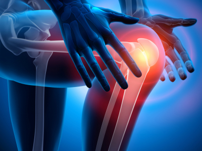
PRP and Knee Pathologies: Indications and Injection Protocol
- Why Does the Knee Respond to PRP?
- Validated Indications at the Knee
- Gonarthrosis
- Intrasubstance Meniscal Lesions
- Tendinopathies
- Focal Cartilage Lesions
- PRP vs Hyaluronic Acid: Comparative Data
- Key Points of the Comparison
- Post-Injection Follow-Up: Key Points for the Practitioner
- Immediate Phase (D0-D3)
- Follow-Up Phase (D3-D30)
The peri-articular tissues of the knee - cartilage, tendons, ligaments, menisci - have limited native vascularisation, which naturally slows their repair capacity. Local PRP injection aims to compensate for this deficit by delivering a high concentration of growth factors directly to the lesion site.
Why Does the Knee Respond to PRP?
Articular cartilage and tendino-ligamentous structures are avascular or hypovascular tissues. In the absence of adequate blood supply, repair cells (fibroblasts, chondrocytes) struggle to migrate to the injured area. PRP injection bypasses this limitation by locally delivering:
- PDGF: proliferation of chondrocytes and fibroblasts
- TGF-b: type II collagen synthesis, inflammation regulation
- VEGF: local neovascularisation
- IGF-1: chondrocyte differentiation and matrix synthesis
- Fibronectin and vitronectin: cell adhesion to extracellular matrix, migration of repair cells
Validated Indications at the Knee
Gonarthrosis
Knee osteoarthritis affects approximately 30% of patients aged 65 to 75 in France. Several prospective studies document a reduction in pain scores (VAS, WOMAC) and functional improvement after PRP injection, with an effect maintained over 6 to 12 months depending on the protocol. A study published in the Official Journal of the Malaysian Orthopaedic Association (2014) reports favourable results for PRP compared to hyaluronic acid for this indication.
Intrasubstance Meniscal Lesions
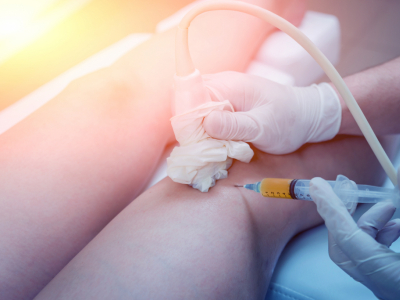
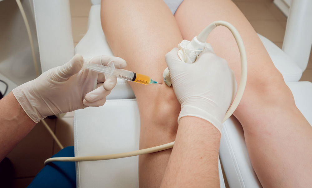
Grade I and II meniscal degenerations in the avascular zone are a documented indication. Ultrasound-guided injections are reported in the literature for these lesions inaccessible to standard minimally invasive surgery.
Tendinopathies
Patellar tendinopathy and quadriceps tendinopathy are frequent indications, particularly in athletes. PRP is used in peri- or intra-tendinous injection under ultrasound guidance to target areas of pathological neovascularisation.
Focal Cartilage Lesions
Focal chondropathies of grade I to III (ICRS classification) represent an indication under evaluation. PRP can be used alone or in combination with other techniques (microfractures, cartilage grafting) to optimise tissue regeneration.
PRP vs Hyaluronic Acid: Comparative Data
The two products have different targets: hyaluronic acid acts primarily as a joint lubricant (viscosupplementation), while PRP targets tissue regeneration through direct biological action. Several meta-analyses report superiority of PRP over hyaluronic acid in pain reduction at medium term (6 months) for mild to moderate gonarthrosis. Results are more homogeneous for leucocyte-poor PRP than for leucocyte-rich PRP for this indication.
Key Points of the Comparison
- PRP: biological action on cartilage matrix, effect at 6-12 months
- Hyaluronic acid: mechanical and anti-inflammatory effect, effect at 3-6 months
- Combination of both possible according to practitioner protocol
- Leucocyte-poor PRP preferred intra-articularly to limit initial inflammatory reaction
Post-Injection Follow-Up: Key Points for the Practitioner
Immediate Phase (D0-D3)
- Local inflammatory reaction possible within 24-48h: normal and expected
- Local cold application in case of swelling
- Partial unloading recommended depending on indication
- Avoid NSAIDs (ibuprofen, diclofenac) which inhibit platelet activation and may reduce PRP efficacy
Follow-Up Phase (D3-D30)
- Grade 1 analgesics (paracetamol) if necessary
- Progressive return to weight-bearing according to indication and tolerance
- Clinical assessment at 4-6 weeks to evaluate initial response
- Multi-injection protocol possible (2 to 3 injections spaced 3-4 weeks apart) depending on practice and indication
Share this content
These products might interest you